Right Bundle Branch Block (RBBB) on the ECG
Right bundle branch block is a conduction delay in the right bundle branch, producing a QRS duration of 120 milliseconds or more with an RSR' pattern in lead V1 and a broad slurred S wave in leads I and V6.
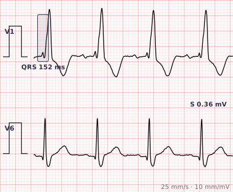
ECG criteria
| Feature | What you see in RBBB |
|---|---|
| QRS | 120 ms or more. 110–120 ms is incomplete RBBB. |
| V1–V2 | RSR' pattern — the classic "M" shape or rabbit ears. Sometimes a broad notched R wave instead. |
| I, V5–V6 | Broad slurred S wave, wider than it is deep. |
| ST-T | ST depression and T wave inversion in V1–V3, discordant to the terminal QRS. Expected, not ischaemic. |
| Axis | Usually normal. A right or left axis alongside RBBB suggests an additional fascicular block. |
| Rate/rhythm | Unaffected — RBBB describes conduction, not rhythm. |
How to spot it
- Measure the QRS. If it is under 120 ms this is not a complete bundle branch block.
- Go to V1. Look for the RSR' — two upward deflections making an M shape. This is the signature.
- Confirm in I and V6 with a broad slurred S wave. Both halves should agree before you call it.
- Check the terminal forces are late and rightward — in RBBB the delay is at the end of the QRS, not the beginning.
- Note the expected ST-T changes in V1–V3 so you do not report them as ischaemia.
- Check the axis. Deviation on top of RBBB means a fascicular block as well, which is bifascicular block.
What it gets confused with
| Looks like | How to tell them apart |
|---|---|
| Left bundle branch block | Broad monophasic R in V6 and a deep S in V1 — the mirror image. LBBB is the more serious of the two. |
| Brugada pattern | Coved ST elevation in V1–V2 with an RBBB-like appearance, but the ST segment is raised and downsloping rather than simply discordant. |
| Posterior myocardial infarction | Tall R in V1 without QRS widening, plus ST depression in V1–V3. |
| Right ventricular hypertrophy | Tall R in V1 with right axis deviation, but a normal QRS duration. |
| Incomplete RBBB | The same morphology with QRS between 110 and 120 ms. Often a normal variant. |
Traps
- The ST depression and T inversion in V1–V3 are part of the block. Reporting them as anterior ischaemia is one of the most common ECG errors. They are expected and should be discordant to the terminal QRS.
- RBBB does not prevent the diagnosis of myocardial infarction the way LBBB traditionally has — Q waves and ST elevation remain interpretable in most territories.
- Isolated RBBB is a common finding in healthy people and is far less likely to indicate structural disease than LBBB.
- New RBBB in a breathless patient should raise the possibility of pulmonary embolism, alongside right heart strain and the S1Q3T3 pattern.
- RBBB plus left anterior fascicular block is bifascicular block; adding a long PR has traditionally been called trifascicular block and warrants attention.
Why it happens
The right bundle branch conducts slowly or not at all, so the right ventricle is not activated through its normal fast pathway. The left ventricle depolarises first and normally, then the impulse crosses the septum and spreads slowly through right ventricular muscle. That delayed rightward activation produces the late R' in V1 and the broad terminal S wave in the left-facing leads.
Why it matters
RBBB is frequently benign and is found in people with structurally normal hearts, particularly as an isolated incidental finding. It gains importance when it is new, when it accompanies fascicular block, or when it appears in an acutely unwell patient — where it may signal pulmonary embolism, right heart strain or extensive conduction system disease.
Questions
What does RBBB look like on an ECG?
A QRS of 120 milliseconds or more, an RSR' pattern in V1 producing an M shape, and a broad slurred S wave in leads I and V6. The ST segment and T wave in V1 to V3 are typically discordant.
Are the T wave changes in RBBB a sign of ischaemia?
No. T wave inversion and ST depression in V1 to V3 are expected consequences of the abnormal repolarisation sequence and should be discordant to the terminal QRS. Concordant changes, or changes in other territories, are what warrant concern.
Is right bundle branch block serious?
Isolated RBBB is common and often benign, particularly in younger people with otherwise normal hearts. It matters more when new, when combined with fascicular block, or when it appears in an acutely unwell patient.
What is the difference between complete and incomplete RBBB?
Only the QRS duration. Complete RBBB has a QRS of 120 milliseconds or more; incomplete RBBB shows the same morphology with a QRS between 110 and 120 milliseconds and is frequently a normal variant.
References
Reading about right bundle branch block is not the same as calling it on a tracing you have never seen.
Practise on real cases in ECG Pro