Left Bundle Branch Block (LBBB) on the ECG
Left bundle branch block is a conduction delay in the left bundle branch, producing a QRS duration of 120 milliseconds or more with a broad notched R wave in leads I, aVL and V6, and a deep S wave or QS complex in V1.
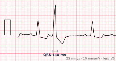
ECG criteria
| Feature | What you see in LBBB |
|---|---|
| QRS | 120 ms or more. 110–120 ms is incomplete LBBB. |
| V1–V2 | Deep broad S wave or QS complex. Usually no R wave at all. |
| I, aVL, V6 | Broad monophasic R wave, often notched or with a plateau at its peak. |
| Q waves | Absent in I, aVL and V6. Septal depolarisation is reversed, so the normal septal Q disappears. |
| ST-T | Discordant — ST elevation with upright T waves in V1–V3, ST depression with T inversion in I, aVL, V5–V6. Expected, not ischaemic. |
| Axis | Usually normal or left. Marked left axis suggests additional disease. |
How to spot it
- Measure the QRS. 120 ms or more is required.
- Go to V1. A deep, broad S wave or a QS complex with no R at all fits LBBB.
- Go to V6 and lead I. A broad monophasic R wave, often notched at the top, confirms it.
- Check the septal Q waves are gone from I, aVL and V6. Their absence is characteristic.
- Note the discordant ST-T changes and do not mistake them for ischaemia.
- If you are assessing for infarction, apply the Sgarbossa criteria rather than reading the ST segments directly.
What it gets confused with
| Looks like | How to tell them apart |
|---|---|
| Right bundle branch block | RSR' in V1 and a broad S in V6 — the mirror pattern. |
| Paced rhythm | Ventricular pacing from the right ventricle produces an LBBB-like pattern. Look for pacing spikes. |
| Ventricular ectopy or VT | Wide complexes without a preceding P wave. LBBB complexes follow a P wave normally. |
| WPW with a left-sided pathway | Short PR and a delta wave slurring the QRS upstroke. LBBB has a normal PR and a sharp initial deflection. |
| Hyperkalaemia | Diffuse QRS widening in all leads with peaked T waves, not a specific bundle branch pattern. |
Traps
- LBBB obscures the usual ECG signs of myocardial infarction. Q waves, ST elevation and T inversion are all altered by the block itself. Use the Sgarbossa criteria — concordant ST elevation of 1 mm or more, concordant ST depression of 1 mm or more in V1–V3, or excessively discordant ST elevation.
- The discordant ST elevation in V1–V3 is expected. Reporting it as an anterior STEMI is a common and consequential error.
- New LBBB is more significant than RBBB and warrants investigation for underlying structural heart disease, even in the absence of symptoms.
- LBBB makes the axis and any assessment of hypertrophy unreliable — the whole depolarisation sequence is abnormal.
- A right ventricular paced rhythm looks very like LBBB. Check for pacing spikes before attributing the pattern to intrinsic conduction disease.
Why it happens
The left bundle branch fails to conduct, so the left ventricle cannot be activated through its normal fast pathway. The septum depolarises from right to left instead of left to right, abolishing the normal septal Q waves, and the left ventricular free wall is then activated slowly through muscle from the right side. That prolonged leftward activation produces the broad monophasic R in the left-facing leads and the deep S in V1.
Why it matters
Left bundle branch block is far more likely than RBBB to indicate underlying structural heart disease — hypertension, coronary disease, aortic valve disease or cardiomyopathy. Its greatest practical importance is that it conceals the ECG changes of infarction, which is why it demands a different approach to chest pain rather than a simple reading of the ST segments.
Questions
What does LBBB look like on an ECG?
A QRS of 120 milliseconds or more, a broad notched monophasic R wave in leads I, aVL and V6, a deep S wave or QS complex in V1, and absent septal Q waves in the lateral leads.
Why does LBBB make myocardial infarction hard to diagnose?
The block already alters the QRS, ST segment and T wave, so the usual signs of infarction are either mimicked or masked. The Sgarbossa criteria were developed to identify infarction despite the block, using concordant ST changes and excessive discordance.
Is new LBBB an emergency?
New LBBB with ischaemic symptoms is treated seriously and has historically been managed like a STEMI equivalent, though current practice relies on the Sgarbossa criteria and the clinical picture rather than the block alone.
Why do septal Q waves disappear in LBBB?
Normally the septum depolarises left to right, producing a small Q wave in the left-facing leads. In LBBB the septum is activated from the right instead, so that initial rightward vector is lost and the Q waves disappear.
References
- Surawicz B et al. AHA/ACCF/HRS Recommendations for the Standardization and Interpretation of the Electrocardiogram, Part III: Intraventricular Conduction Disturbances. Circulation 2009.
- Sgarbossa EB et al. Electrocardiographic diagnosis of evolving acute myocardial infarction in the presence of left bundle-branch block. N Engl J Med 1996.
Reading about left bundle branch block is not the same as calling it on a tracing you have never seen.
Practise on real cases in ECG Pro