S1Q3T3 on the ECG
S1Q3T3 is an ECG pattern consisting of a prominent S wave in lead I, a Q wave in lead III and an inverted T wave in lead III, classically associated with acute pulmonary embolism but present in only a minority of cases.
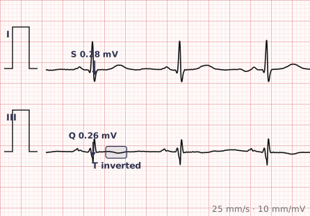
ECG criteria
| Feature | What you see in S1Q3T3 |
|---|---|
| Lead I | Prominent S wave, usually at least 1.5 mm. |
| Lead III | Q wave, usually at least 1.5 mm. |
| Lead III | Inverted T wave. |
| Sensitivity | Low. Present in roughly a fifth of confirmed pulmonary emboli, so its absence excludes nothing. |
| More common findings | Sinus tachycardia is the most common ECG finding in PE. T inversion in V1–V4 is more sensitive than S1Q3T3. |
| Other signs | Right axis deviation, right bundle branch block, right atrial enlargement, atrial fibrillation. |
How to spot it
- Look at lead I for a prominent S wave.
- Look at lead III for both a Q wave and an inverted T wave. All three components are needed.
- Recognise immediately that the pattern is neither sensitive nor specific — it supports the diagnosis but never makes or excludes it.
- Look for the findings that are actually more common: sinus tachycardia, and T wave inversion in V1 to V4.
- Look for other evidence of right heart strain — right axis deviation, right bundle branch block, right atrial enlargement.
- Return to the clinical picture and a validated probability score. The ECG is supporting evidence in pulmonary embolism, not the test.
What it gets confused with
| Looks like | How to tell them apart |
|---|---|
| Normal variant | A small Q wave and S wave pattern occurs in healthy people, particularly those with a vertical heart. |
| Inferior myocardial infarction | Q waves in II, III and aVF with ST changes, not just lead III. |
| Right ventricular hypertrophy | Dominant R in V1 with right axis deviation, in a chronic rather than acute picture. |
| Chronic lung disease | Low voltage, right axis deviation and right atrial enlargement, developing gradually. |
| Left posterior fascicular block | Right axis deviation with rS in I and qR in III. |
Traps
- S1Q3T3 is far better known than it is useful. It appears in only around a fifth of pulmonary emboli, and it also occurs in healthy people. It cannot rule the diagnosis in or out.
- Sinus tachycardia is the most common ECG finding in pulmonary embolism, and a normal ECG is entirely compatible with a large embolus.
- T wave inversion in V1 to V4 is more sensitive for pulmonary embolism than S1Q3T3 and is more useful to look for.
- A Q wave in lead III alone is a common normal finding — all three components must be present before the pattern is named.
- Never let the absence of S1Q3T3 reduce clinical suspicion. The diagnosis rests on probability assessment and imaging.
Why it happens
Acute obstruction of the pulmonary arteries raises right ventricular afterload sharply, causing acute dilatation and strain of a chamber unaccustomed to high pressure. That rotates the heart and shifts the electrical axis rightward and posteriorly, producing the S wave in lead I and the Q wave in lead III, while strain on the right ventricular free wall produces the T wave inversion.
Why it matters
The value of S1Q3T3 lies mostly in knowing how little it proves. It is one of the most frequently taught and least useful ECG patterns, and its main clinical role is as a reminder that pulmonary embolism cannot be excluded by an ECG. Right heart strain findings taken together — tachycardia, anterior T inversion, right axis shift — are more informative than any one pattern.
Questions
How sensitive is S1Q3T3 for pulmonary embolism?
Poorly. It appears in roughly 20 per cent of confirmed pulmonary emboli, so its absence provides no reassurance. It also occurs in healthy people and in other conditions causing right heart strain.
What is the most common ECG finding in pulmonary embolism?
Sinus tachycardia. T wave inversion in leads V1 to V4 is the next most useful finding and is more sensitive than the S1Q3T3 pattern. A completely normal ECG is also entirely compatible with pulmonary embolism.
What does S1Q3T3 actually consist of?
A prominent S wave in lead I, a Q wave in lead III, and an inverted T wave in lead III. All three components must be present for the pattern to be named.
Can S1Q3T3 occur in healthy people?
Yes. A Q wave in lead III is a common normal finding, particularly in people with a vertically oriented heart, and the full pattern can occur without any underlying pathology.
Reading about s1q3t3 is not the same as calling it on a tracing you have never seen.
Practise on real cases in ECG Pro