ST Elevation on the ECG
ST elevation is displacement of the ST segment above the isoelectric baseline, measured at the J point, which may indicate acute myocardial injury but also occurs in pericarditis, early repolarization, left ventricular hypertrophy, bundle branch block and several other conditions.
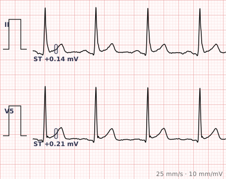
ECG criteria
| Feature | What you see in ST Elevation |
|---|---|
| Threshold | ≥ 1 mm in two contiguous leads, in every lead except V2–V3. In V2–V3: ≥ 2.5 mm (men under 40), ≥ 2 mm (men 40+), ≥ 1.5 mm (women). |
| Shape — convex | Convex upward, or "tombstone". Strongly suggests infarction. |
| Shape — concave | Concave upward. More typical of pericarditis and early repolarization, though it does not exclude infarction. |
| Distribution | Territorial (grouped by coronary supply) suggests infarction. Widespread across territories suggests pericarditis. |
| Reciprocal change | Present in infarction, absent in pericarditis. The most useful single feature. |
| PR segment | PR depression with PR elevation in aVR supports pericarditis. |
How to spot it
- Measure at the J point, taking the TP segment as the baseline.
- Confirm the elevation reaches threshold in two contiguous leads.
- Look at the shape. Convex or straightened elevation favours infarction; concave favours the benign causes.
- Map the distribution. Does it fit a coronary territory, or is it spread across several?
- Look for reciprocal depression in the opposite leads — the single most useful discriminator.
- Check the PR segments. PR depression with elevation in aVR points to pericarditis.
- Then rule out the structural mimics: LVH, LBBB, pacing and Brugada all elevate the ST segment for their own reasons.
What it gets confused with
| Looks like | How to tell them apart |
|---|---|
| STEMI | Territorial, convex, with reciprocal depression and evolving over time. |
| Pericarditis | Widespread and concave, with PR depression and no reciprocal change. |
| Early repolarization | Concave with J point notching, in the young, and stable across tracings. |
| Left ventricular hypertrophy | ST elevation in V1–V3 as a secondary change to deep S waves, with voltage criteria met. |
| Left bundle branch block | Discordant elevation is expected. Apply the Sgarbossa criteria. |
| Brugada pattern | Coved elevation in V1–V2 with a partial RBBB appearance. |
| Ventricular aneurysm | Persistent ST elevation with Q waves, unchanged over months in a patient with a previous infarct. |
Traps
- ST elevation is a finding, not a diagnosis. Reading it as automatically meaning infarction leads to inappropriate reperfusion; assuming it is benign leads to missed occlusion. Shape, distribution and reciprocal change decide.
- Take the TP segment as baseline, not the PR segment, particularly when PR depression is present.
- Persistent ST elevation with established Q waves in a patient with an old infarct suggests a ventricular aneurysm rather than a new event.
- A single tracing cannot distinguish evolving from static change. Serial ECGs are frequently the most informative test available.
- In left bundle branch block or a paced rhythm, ST segments cannot be read conventionally at all.
Why it happens
The ST segment corresponds to the plateau phase, when the whole ventricle should be uniformly depolarised and no potential difference exists. Anything that makes one region electrically different from another during that phase creates a current that displaces the segment — acutely injured myocardium being the classic example, but also inflamed pericardium, hypertrophied muscle and abnormal repolarisation from conduction disease.
Why it matters
ST elevation is the single most consequential pattern on the ECG, because it triggers emergency reperfusion. Its importance lies as much in the discipline of assessing shape, distribution and reciprocal change as in spotting the elevation itself — the difference between a STEMI and pericarditis is the difference between the catheter laboratory and an anti-inflammatory.
Questions
Does ST elevation always mean a heart attack?
No. Pericarditis, early repolarization, left ventricular hypertrophy, left bundle branch block, ventricular pacing, Brugada syndrome and ventricular aneurysm all produce ST elevation. The shape, distribution and presence of reciprocal change determine which.
What is the difference between concave and convex ST elevation?
Convex or straightened elevation, sometimes called tombstone, strongly suggests acute infarction. Concave elevation is more typical of pericarditis and early repolarization, although concave morphology does not exclude infarction.
How do I distinguish pericarditis from a STEMI?
Pericarditis usually produces widespread concave ST elevation across several coronary territories, with PR segment depression and no reciprocal ST depression. A STEMI produces elevation confined to one territory with reciprocal depression in the opposite leads.
Where should ST elevation be measured from?
At the J point, using the TP segment as the baseline. Using the PR segment as baseline is unreliable when PR depression is present, as it is in pericarditis.
References
- Thygesen K et al. Fourth Universal Definition of Myocardial Infarction (2018). J Am Coll Cardiol.
- Rautaharju PM et al. AHA/ACCF/HRS Recommendations for the Standardization and Interpretation of the Electrocardiogram, Part IV: ST Segment, T and U Waves, and the QT Interval. Circulation 2009.
Reading about st elevation is not the same as calling it on a tracing you have never seen.
Practise on real cases in ECG Pro