Right Atrial Enlargement (RAE) on the ECG
Right atrial enlargement on the ECG is indicated by a tall peaked P wave in lead II measuring 2.5 millimetres or more in height, with a normal P wave duration and often a prominent initial positive deflection in lead V1.
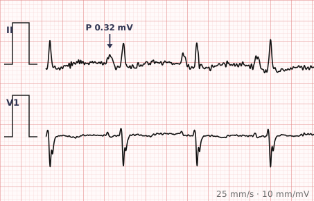
ECG criteria
| Feature | What you see in RAE |
|---|---|
| Lead II | P wave ≥ 2.5 mm tall, peaked and symmetric. Classically "P pulmonale". |
| Lead V1 | Initial positive deflection ≥ 1.5 mm. The early part of the P wave is enlarged. |
| P duration | Normal, under 120 ms. The P is taller, not wider — this separates it from left atrial enlargement. |
| Axis | P wave axis often shifts rightward, towards +75° or beyond. |
| Supporting | Frequently accompanies right ventricular hypertrophy and chronic lung disease. |
| Rhythm | Sinus. Not assessable in atrial fibrillation. |
How to spot it
- Go to lead II and measure the height of the P wave. 2.5 mm or more — two and a half small squares — meets the criterion.
- Check the P wave duration is normal. A P that is both tall and broad suggests biatrial enlargement.
- Look at V1 for a prominent initial positive component.
- Note the P wave shape: right atrial enlargement gives a peaked, symmetric P, not a notched one.
- Look for right ventricular hypertrophy and right axis deviation, which commonly accompany it and point towards the same underlying cause.
What it gets confused with
| Looks like | How to tell them apart |
|---|---|
| Left atrial enlargement | Broad and notched rather than tall and peaked. |
| Biatrial enlargement | P wave both tall and broad, with a large biphasic P in V1. |
| Sinus tachycardia | P waves become taller and more peaked at fast rates without any atrial enlargement. |
| Hypokalaemia | Can increase P wave amplitude, alongside U waves and ST depression. |
| Pulmonary embolism | Acute right heart strain can produce transient peaked P waves with right axis shift. |
Traps
- Check the rate before calling P pulmonale. Sinus tachycardia alone makes P waves taller and more peaked, and a rate of 130 can produce the appearance without any atrial pathology.
- Right atrial enlargement makes the P taller; left atrial enlargement makes it wider. Confusing the two is the most common error.
- The finding is often transient in acute conditions such as pulmonary embolism or an asthma exacerbation, and may resolve entirely on recovery.
- "Right atrial abnormality" is often the more accurate phrase, since the ECG reflects conduction rather than measured chamber size.
- A tall P wave with low QRS voltages and right axis deviation is a common chronic lung disease pattern.
Why it happens
The right atrium depolarises first, so it contributes to the early part of the P wave. When it is enlarged, that early component carries more voltage and points more directly towards the inferior leads, producing a taller peaked P wave in lead II. Because activation of the two atria still overlaps in the normal way, the total P wave duration is unchanged — only its amplitude increases.
Why it matters
Right atrial enlargement points towards conditions that load the right heart: chronic obstructive pulmonary disease, pulmonary hypertension, pulmonary embolism, pulmonary or tricuspid valve disease, and congenital lesions. On the ECG it rarely stands alone, and its main use is as one component of a right heart strain picture.
Questions
What is P pulmonale?
The classic ECG appearance of right atrial enlargement — a tall, peaked P wave in lead II measuring 2.5 millimetres or more, with a normal P wave duration. The name reflects its association with pulmonary disease.
Can a fast heart rate mimic right atrial enlargement?
Yes. Sinus tachycardia increases P wave amplitude and makes P waves appear more peaked, so the finding should always be interpreted with the rate in mind.
How do I distinguish right from left atrial enlargement?
Height versus width. Right atrial enlargement produces a tall peaked P wave of normal duration; left atrial enlargement produces a broad notched P wave lasting 120 milliseconds or more.
What conditions cause right atrial enlargement?
Anything that loads the right heart chronically or acutely — chronic obstructive pulmonary disease, pulmonary hypertension, pulmonary embolism, tricuspid or pulmonary valve disease, and some congenital heart lesions.
References
Reading about right atrial enlargement is not the same as calling it on a tracing you have never seen.
Practise on real cases in ECG Pro