Left Atrial Enlargement (LAE) on the ECG
Left atrial enlargement on the ECG is indicated by a prolonged and often notched P wave in lead II lasting 120 milliseconds or more, together with a deep terminal negative deflection of the P wave in lead V1.
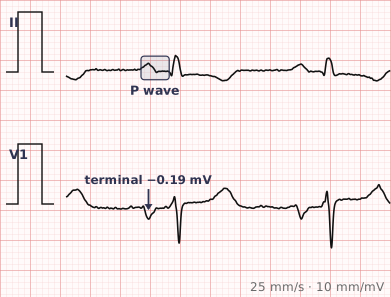
ECG criteria
| Feature | What you see in LAE |
|---|---|
| Lead II | P wave ≥ 120 ms, often bifid with notching and a peak-to-peak interval over 40 ms. Classically "P mitrale". |
| Lead V1 | Deep terminal negative component — at least 1 mm deep and 40 ms wide. Known as the Morris index. |
| P amplitude | Normal. The abnormality is duration and shape, not height. |
| Axis | P wave axis usually normal. |
| Supporting | Frequently accompanies left ventricular hypertrophy, mitral valve disease and hypertension. |
| Rhythm | Sinus. In atrial fibrillation there are no P waves to assess. |
How to spot it
- Go to lead II and measure the P wave duration. 120 ms or more — three small squares — is the threshold.
- Look at the P wave shape in II. Notching with a broad, camel-hump appearance supports enlargement.
- Go to V1 and look at the terminal negative part of the P wave. A deflection 1 mm deep and 40 ms wide is the Morris index and is the more specific sign.
- Check that the P amplitude is normal. Tall peaked P waves point to the right atrium instead.
- Look for the company it keeps — left ventricular hypertrophy and mitral valve disease commonly accompany it.
What it gets confused with
| Looks like | How to tell them apart |
|---|---|
| Right atrial enlargement | Tall and peaked P wave rather than broad and notched. Amplitude versus duration is the whole distinction. |
| Biatrial enlargement | Both features present — a P wave that is tall and broad, with a large biphasic P in V1. |
| Interatrial block | P wave ≥ 120 ms with a biphasic P in the inferior leads. Overlaps with LAE and is increasingly reported separately. |
| Artefact or baseline wander | Apparent P wave broadening from a wandering baseline. Check across several beats. |
Traps
- The distinction from right atrial enlargement is duration versus amplitude. Left atrial enlargement makes the P wave wider; right atrial enlargement makes it taller. Learning it that way prevents most errors.
- The V1 terminal negativity — the Morris index — is more specific than the lead II duration, so check both.
- "Left atrial abnormality" is often preferred to "enlargement", since the ECG changes reflect conduction through the atrium rather than measured size.
- P wave criteria cannot be applied in atrial fibrillation, where there are no P waves at all.
- Left atrial enlargement is common in hypertension and mitral valve disease, and is one of the ECG markers associated with developing atrial fibrillation.
Why it happens
Depolarisation of the atria proceeds from right to left. When the left atrium is enlarged or conducts slowly, the leftward component of atrial activation takes longer and finishes later, which lengthens the overall P wave and separates its two humps in lead II. Because the enlarged left atrium sits posteriorly, the terminal part of that activation moves away from V1, producing the deep terminal negative deflection there.
Why it matters
Left atrial enlargement reflects chronic pressure or volume loading of the left atrium, most often from hypertension, left ventricular hypertrophy, or mitral valve disease. Its practical value is as a marker of chronicity and as a predictor: the enlarged, slowly conducting left atrium is the substrate on which atrial fibrillation develops.
Questions
What is P mitrale?
The classic ECG appearance of left atrial enlargement — a broad, notched, bifid P wave in lead II lasting 120 milliseconds or more. The name reflects its historical association with mitral valve disease.
What is the Morris index?
The terminal negative portion of the P wave in lead V1, measured as depth multiplied by duration. A terminal deflection at least 1 millimetre deep and 40 milliseconds wide is a specific sign of left atrial enlargement.
How do I tell left from right atrial enlargement?
Left atrial enlargement broadens the P wave; right atrial enlargement heightens it. A P wave in lead II that is wide and notched points left, one that is tall and peaked points right.
Why is it sometimes called left atrial abnormality?
Because the ECG changes reflect the pattern and timing of atrial conduction rather than a direct measurement of chamber size. The term abnormality avoids implying a size that the ECG cannot actually measure.
References
Reading about left atrial enlargement is not the same as calling it on a tracing you have never seen.
Practise on real cases in ECG Pro