Pulmonary Heart Disease on the ECG
Pulmonary heart disease, or cor pulmonale, is right ventricular enlargement and dysfunction caused by disease of the lungs or pulmonary circulation, producing an ECG pattern of right atrial enlargement, right axis deviation, right ventricular hypertrophy and often low voltage.
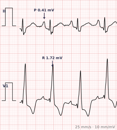
ECG criteria
| Feature | What you see in Pulmonary Heart Disease |
|---|---|
| P waves | Tall peaked P waves ≥ 2.5 mm in lead II — P pulmonale, from right atrial enlargement. |
| Axis | Right axis deviation, often with a vertical or rightward P wave axis too. |
| V1 | Dominant R wave if right ventricular hypertrophy has developed. |
| Voltage | Low voltage in the limb leads, from hyperinflated lungs insulating the heart. |
| R wave progression | Poor, with a delayed transition, from rotation and hyperinflation. |
| Rhythm | Sinus tachycardia common. Multifocal atrial tachycardia is characteristic of severe COPD. |
How to spot it
- Look at lead II for tall peaked P waves — right atrial enlargement is often the earliest and most visible sign.
- Check the axis for right deviation.
- Look at V1 for a dominant R wave, indicating right ventricular hypertrophy.
- Assess overall voltage. Low limb lead voltage with poor R wave progression fits a hyperinflated chest.
- Check the rhythm. Multifocal atrial tachycardia — three or more P wave shapes at over 100 — is strongly associated with severe lung disease.
- Read the pattern as a whole. No single feature is diagnostic; the combination is what points to the right heart.
What it gets confused with
| Looks like | How to tell them apart |
|---|---|
| Acute pulmonary embolism | Sudden onset with sinus tachycardia and anterior T inversion, sometimes S1Q3T3. Cor pulmonale develops gradually. |
| Right ventricular hypertrophy from other causes | Congenital heart disease or pulmonary stenosis rather than lung disease. |
| Posterior myocardial infarction | Tall R in V1 with ST depression in V1–V3 and a normal axis. |
| Pericardial effusion | Low voltage with electrical alternans and sinus tachycardia, without right atrial enlargement. |
| Obesity | Low voltage without right heart features. |
Traps
- No single ECG feature diagnoses cor pulmonale. It is a pattern recognised from the combination of right atrial enlargement, right axis deviation and low voltage, and the ECG is insensitive early in the disease.
- Low voltage in chronic lung disease comes from hyperinflated lungs insulating the heart, and it can mask right ventricular hypertrophy that is genuinely present.
- Multifocal atrial tachycardia is highly characteristic of severe COPD and is frequently misread as atrial fibrillation. Look for the multiple distinct P wave shapes.
- The ECG findings are those of chronic right heart strain, so they do not distinguish the underlying lung disease. That requires imaging and lung function testing.
- Acute pulmonary embolism and chronic cor pulmonale can look similar on a single tracing. The timing and clinical picture separate them.
Why it happens
Chronic lung disease raises pulmonary vascular resistance through hypoxic vasoconstriction and destruction of the pulmonary vascular bed. The right ventricle faces sustained pressure overload, hypertrophies and eventually dilates, shifting the electrical axis rightward and increasing right-sided voltages. Hyperinflated lungs simultaneously act as an insulator between heart and electrodes, which is why the tracing often shows low voltage despite genuine hypertrophy.
Why it matters
The ECG in pulmonary heart disease is a window on the right side of the circulation, and its findings mark disease that is already well established. Recognising the combination matters because it reframes a breathless patient — a low-voltage tracing with peaked P waves and right axis deviation points towards the lungs and the right heart rather than towards left ventricular failure or coronary disease.
Questions
What is cor pulmonale?
Right ventricular enlargement and dysfunction caused by disease of the lungs or pulmonary circulation, most commonly chronic obstructive pulmonary disease. It reflects sustained pressure overload of the right ventricle.
What are the ECG features of chronic lung disease?
Tall peaked P waves in lead II from right atrial enlargement, right axis deviation, low voltage in the limb leads, poor R wave progression, and sometimes a dominant R wave in V1 from right ventricular hypertrophy.
Why is the ECG voltage low in chronic lung disease?
Hyperinflated lungs contain more air between the heart and the chest electrodes, and air is a poor conductor. The insulating effect reduces the recorded amplitude of all complexes, and can mask genuine right ventricular hypertrophy.
What is multifocal atrial tachycardia and why does it matter here?
An irregular rhythm above 100 beats per minute with at least three different P wave morphologies. It is strongly associated with severe chronic obstructive pulmonary disease and is frequently misdiagnosed as atrial fibrillation, which has no P waves at all.
Reading about pulmonary heart disease is not the same as calling it on a tracing you have never seen.
Practise on real cases in ECG Pro