ST-T Change on the ECG
ST-T change is a general term for any abnormality of the ST segment or T wave that does not meet criteria for a more specific diagnosis, and it is among the most frequently reported findings on routine ECGs.
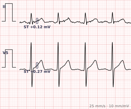
ECG criteria
| Feature | What you see in ST-T Change |
|---|---|
| ST segment | Depression or elevation below the thresholds for a specific diagnosis, often under 1 mm and without a clear territorial pattern. |
| T wave | Flattening, low amplitude, biphasic shape, or inversion in leads where inversion is not expected. |
| Distribution | Often scattered rather than territorial. A pattern confined to one coronary territory demands a more specific label. |
| Reciprocal change | Absent. Its presence would point to infarction instead. |
| Evolution | Static across serial tracings. Change over hours moves the finding into acute territory. |
| Context | Frequently secondary to hypertrophy, bundle branch block, drugs, electrolytes or rate. |
How to spot it
- Establish that there is a genuine ST or T abnormality, measuring at the J point against the TP baseline.
- Ask whether it meets criteria for anything specific — STEMI, a strain pattern, pericarditis, digoxin effect. If it does, use that name instead.
- Map the distribution. Territorial grouping is a reason to look harder; scattered changes are more likely to be non-specific.
- Look for a secondary cause. Hypertrophy, bundle branch block, pre-excitation and pacing all produce expected repolarisation changes that should not be reported as primary.
- Check rate, drugs and electrolytes — all three produce ST-T change that resolves when corrected.
- Compare with a previous ECG. Static change over years is reassuring in a way no single tracing can be.
What it gets confused with
| Looks like | How to tell them apart |
|---|---|
| Myocardial ischaemia | Territorial, horizontal or downsloping depression, symmetric T inversion, and evolving. |
| LVH strain | Downsloping depression with asymmetric T inversion in leads with tall R waves, and voltage criteria met. |
| Bundle branch block | Discordant changes expected as a secondary consequence of abnormal depolarisation. |
| Digoxin effect | Scooped, reverse-tick ST depression with a short QT. |
| Electrolyte disturbance | Hypokalaemia gives flat T waves with prominent U waves; hyperkalaemia gives peaked T waves. |
| Rate-related change | Appears with tachycardia and resolves as the rate settles. |
Traps
- "Non-specific ST-T change" is a description of uncertainty, not a diagnosis. It is appropriate when nothing more specific fits, and inappropriate as a way of avoiding a diagnosis that does.
- It is extremely common — the keyword matches 384 of the 1,389 cases in the ECG Pro library — which is precisely why it must not be used to dismiss a symptomatic patient.
- Secondary changes from hypertrophy, bundle branch block or pacing are expected and should be named as such rather than reported as non-specific.
- The single most useful next step is a previous ECG. New change in a symptomatic patient is a different finding from the same change present for a decade.
- Serial tracings matter more here than almost anywhere else: evolution is what separates non-specific from acute.
Why it happens
The ST segment and T wave represent ventricular repolarisation, a process sensitive to almost every influence on the myocardium — perfusion, wall thickness, activation sequence, membrane potential, autonomic tone, drugs and electrolytes. Because so many mechanisms converge on the same part of the waveform, the resulting abnormality frequently carries no signature identifying which one caused it.
Why it matters
ST-T change is the most common abnormal finding on routine ECGs and the one most often over- and under-interpreted. Reported carefully it is honest: something is different about repolarisation, and the ECG cannot say what. Reported carelessly it becomes either false reassurance for a patient with real ischaemia, or an unnecessary cascade of investigation in someone well.
Questions
What does non-specific ST-T change mean?
It means the ST segment or T wave is abnormal, but the abnormality does not meet criteria for any specific diagnosis. It is a statement of uncertainty rather than a diagnosis in itself.
Is ST-T change serious?
It depends entirely on context. In an asymptomatic patient with an identical tracing from years earlier it usually means little. In someone with chest pain it cannot be used as reassurance, and warrants serial tracings and clinical assessment.
What causes non-specific ST-T changes?
Ventricular hypertrophy, bundle branch block, ventricular pacing, drugs including digoxin, electrolyte disturbances, fast heart rates, and myocardial ischaemia that has not yet declared itself in a specific pattern.
What should I do when I see ST-T change?
Look for a more specific pattern first, look for a secondary cause such as hypertrophy or a conduction abnormality, and then compare with any previous ECG. A change that is new carries far more weight than one that has been present for years.
References
Reading about st-t change is not the same as calling it on a tracing you have never seen.
Practise on real cases in ECG Pro