Right Axis Deviation (RAD) on the ECG
Right axis deviation is a mean QRS axis between +90 and +180 degrees, recognised on the ECG by a predominantly negative QRS complex in lead I with a predominantly positive QRS in lead aVF.
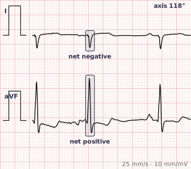
ECG criteria
| Feature | What you see in RAD |
|---|---|
| Axis | +90° to +180°. Beyond +180° is extreme axis deviation, the "northwest" quadrant. |
| Lead I | Negative. |
| Lead aVF | Positive. |
| Lead III | Tall R wave, usually the most positive of the limb leads. |
| QRS | Duration normal unless there is coexisting block. |
| Supporting | Look for right ventricular hypertrophy and right atrial enlargement. |
How to spot it
- Look at leads I and aVF. Lead I negative with aVF positive places the axis in the right quadrant.
- Confirm with lead III, which should show a tall dominant R wave.
- Check V1 for a dominant R wave, which would support right ventricular hypertrophy as the cause.
- Check lead II for tall peaked P waves suggesting right atrial enlargement.
- Consider the patient's age and build — right axis deviation is normal in children and common in tall, thin adults.
- Check lead placement. Reversed arm leads produce a spurious axis shift and are a classic trap.
What it gets confused with
| Looks like | How to tell them apart |
|---|---|
| Normal in children | Right axis deviation is normal in neonates and young children. |
| Right ventricular hypertrophy | Dominant R in V1 with right atrial enlargement and often a strain pattern. |
| Lateral myocardial infarction | Q waves in I and aVL remove the leftward forces and shift the axis right. |
| Left posterior fascicular block | Right axis with rS in I and qR in III, and no other cause. A diagnosis of exclusion. |
| Pulmonary embolism | Acute right heart strain, often with S1Q3T3 and sinus tachycardia. |
| Lead reversal | Arm lead reversal inverts lead I and mimics right axis deviation. Check the P wave in aVR. |
Traps
- Check for limb lead reversal before accepting right axis deviation. Reversed arm leads invert lead I and produce a convincing but false axis shift. A positive P wave in aVR is the giveaway.
- Right axis deviation is normal in neonates and young children, where the right ventricle is still relatively dominant.
- It is also common in tall, thin adults with a vertically oriented heart, and needs no investigation on its own.
- Left posterior fascicular block is rare and should only be diagnosed once the common causes have been excluded.
- In an acutely breathless patient, new right axis deviation should raise the possibility of pulmonary embolism.
Why it happens
The mean QRS axis swings rightward whenever right ventricular forces increase relative to left, or left-sided forces are lost. Right ventricular hypertrophy adds mass on the right; lateral infarction removes myocardium on the left; and in children the right ventricle is naturally more prominent, all producing a vector that points down and to the right.
Why it matters
In an adult, right axis deviation usually points towards the right heart and the pulmonary circulation, and its main value is prompting a look at V1, the P waves and the clinical picture. In children it is a normal developmental finding, which makes age an essential part of interpreting it — one of the clearest examples of why paediatric and adult ECGs cannot be read by the same rules.
Questions
How do I recognise right axis deviation?
A predominantly negative QRS complex in lead I with a predominantly positive QRS in lead aVF. Lead III usually shows the tallest R wave of the limb leads.
Is right axis deviation normal in children?
Yes. The right ventricle is relatively dominant at birth and for the first years of life, so right axis deviation is an expected finding that gradually resolves as the left ventricle becomes dominant.
What is extreme axis deviation?
A mean QRS axis between −90 and ±180 degrees, sometimes called the northwest quadrant, with both lead I and lead aVF negative. It is uncommon and is seen with ventricular rhythms, hyperkalaemia and some congenital conditions.
Can lead reversal mimic right axis deviation?
Yes, and it is a common trap. Reversal of the arm electrodes inverts lead I and produces an apparent right axis shift. A positive P wave in aVR, which should normally be negative, indicates the leads are reversed.
Reading about right axis deviation is not the same as calling it on a tracing you have never seen.
Practise on real cases in ECG Pro