Left Axis Deviation (LAD) on the ECG
Left axis deviation is a mean QRS axis between −30 and −90 degrees, recognised on the ECG by a predominantly positive QRS complex in lead I with a predominantly negative QRS in leads II and aVF.
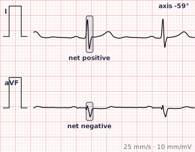
ECG criteria
| Feature | What you see in LAD |
|---|---|
| Axis | −30° to −90°. Beyond −45° suggests left anterior fascicular block. |
| Lead I | Positive. |
| Lead aVF | Negative. |
| Lead II | Negative confirms the axis is beyond −30°. This is the quick bedside test. |
| QRS | Duration depends on the cause — normal in fascicular block, wide in bundle branch block. |
| Other leads | aVL typically shows a tall R wave. |
How to spot it
- Look at leads I and aVF. Lead I positive with aVF negative places the axis in the left quadrant.
- Then look at lead II. If lead II is also predominantly negative, the axis is beyond −30° and this is genuine left axis deviation rather than a normal leftward axis.
- Measure the QRS width. Narrow with marked left axis suggests left anterior fascicular block; wide suggests bundle branch block.
- Look at the inferior leads for pathological Q waves, since previous inferior infarction shifts the axis leftward.
- Check the voltage criteria for left ventricular hypertrophy, another common cause.
- Report the likely reason rather than the axis alone — "left axis deviation due to left anterior fascicular block" is a useful statement; "left axis deviation" on its own is not.
What it gets confused with
| Looks like | How to tell them apart |
|---|---|
| Left anterior fascicular block | Axis −45° or beyond, qR in aVL, rS inferiorly, narrow QRS. The most common cause. |
| Inferior myocardial infarction | Pathological Q waves in II, III and aVF. |
| Left ventricular hypertrophy | Voltage criteria met, often with a lateral strain pattern. |
| Left bundle branch block | QRS 120 ms or more with a broad monophasic R in V6. |
| WPW | Short PR with a delta wave; pre-excitation can shift the axis in any direction. |
| Normal variant | Axis between 0 and −30°, common with obesity, pregnancy and increasing age. |
Traps
- Lead II is the fastest discriminator. A negative lead II means the axis is beyond −30° and the leftward shift is genuine rather than a normal variant.
- An axis between 0 and −30° is common and normal, particularly in older, heavier or pregnant patients — it does not need reporting as an abnormality.
- Left axis deviation is a finding, not a diagnosis. The value is in identifying the cause.
- Fascicular block and inferior infarction both shift the axis left and are distinguished by whether the inferior leads show an rS or a Q wave.
- A wide QRS makes axis interpretation less reliable, since the activation sequence is already abnormal.
Why it happens
The mean QRS axis represents the average direction of ventricular depolarisation. It swings leftward and superiorly whenever activation of the left and superior parts of the ventricle is delayed relative to the rest — as in left anterior fascicular block — or whenever inferior myocardium is lost to infarction, removing the downward vector that normally balances it.
Why it matters
Left axis deviation is common and frequently benign, but it is a useful gateway finding: it prompts a check for fascicular block, previous inferior infarction and left ventricular hypertrophy, each of which carries its own significance. Its greatest value is that it costs two seconds to notice and directs where to look next.
Questions
How do I recognise left axis deviation?
A predominantly positive QRS in lead I with a predominantly negative QRS in lead aVF. If lead II is also negative, the axis is beyond −30 degrees and the deviation is genuine.
What is the most common cause of left axis deviation?
Left anterior fascicular block, in which the anterior fascicle of the left bundle branch fails to conduct and the ventricle is activated in a sequence that swings the axis sharply leftward.
Is left axis deviation always abnormal?
No. An axis between 0 and −30 degrees is a normal variant and is common with increasing age, obesity and pregnancy. Deviation beyond −30 degrees is more likely to reflect an underlying cause.
Reading about left axis deviation is not the same as calling it on a tracing you have never seen.
Practise on real cases in ECG Pro