Clockwise Rotation on the ECG
Clockwise rotation is a shift of the precordial R/S transition point later than normal, towards leads V5 and V6, reflecting rotation of the heart about its long axis as viewed from below the diaphragm.
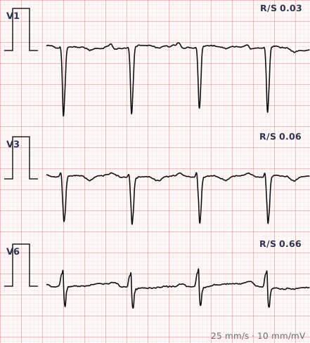
ECG criteria
| Feature | What you see in Clockwise Rotation |
|---|---|
| Transition | R/S transition at V5 or later, instead of the usual V3–V4. |
| V1–V4 | Persistent S wave dominance, with small R waves that grow late. |
| V5–V6 | The transition finally occurs here, sometimes not at all. |
| QRS | Normal duration. Widening points to a conduction abnormality instead. |
| Axis | Often normal or rightward, particularly with lung disease. |
| Associated | Chronic lung disease, right ventricular enlargement, and a vertical heart in tall thin people. |
How to spot it
- Scan V1 to V6 and find the lead where the R wave first exceeds the S wave. That is the transition point.
- Normal transition sits at V3 or V4. At V5 or later, the rotation is clockwise.
- Check the QRS width, to make sure a conduction abnormality is not producing the appearance.
- Check lead placement. Electrodes too high shift the transition and are a common cause.
- Look for supporting features of lung disease or right heart strain — low voltage, right axis deviation, peaked P waves.
- Report it as a descriptive finding, not a diagnosis. It says how the heart is oriented, not what is wrong with it.
What it gets confused with
| Looks like | How to tell them apart |
|---|---|
| Poor R wave progression | Overlapping and often used interchangeably. PRWP is defined by R wave amplitude in V3; rotation is defined by where the transition falls. |
| Anterior myocardial infarction | Pathological Q waves in the anterior leads, often with T inversion. |
| Counter-clockwise rotation | The opposite — transition early, at V2 or before. |
| Chronic lung disease | Low voltage, right axis deviation, peaked P waves alongside the late transition. |
| Lead misplacement | Chest electrodes an intercostal space too high. Repeat with careful placement. |
| Left bundle branch block | Wide QRS with a broad R in V6; the transition is displaced as part of the block. |
Traps
- Clockwise rotation is a description of position, not a diagnosis. Reporting it as though it means disease over-reads a finding that is frequently anatomical.
- It overlaps heavily with poor R wave progression, and the two terms are often used for the same tracing. Being explicit about which definition you are applying avoids confusion.
- Lead placement is the most common artefactual cause, exactly as it is for poor R wave progression.
- It is common in chronic lung disease and in tall thin people with a vertically hanging heart, in neither of whom does it require investigation on its own.
- The name describes the heart viewed from below the diaphragm looking up — which is why the direction feels counter-intuitive when first learned.
Why it happens
The heart can rotate about its long axis, the axis running from base to apex. Viewed from below the diaphragm, clockwise rotation swings the right ventricle further leftward and anteriorly, so the transition from right ventricular to left ventricular dominance occurs later across the chest leads. Hyperinflated lungs and a vertically oriented heart both promote it, which is why it accompanies chronic lung disease so often.
Why it matters
Clockwise rotation is a positional descriptor with modest independent weight. Its practical value is in preventing over-reading: a late transition explains small anterior R waves without requiring an infarct, and recognising it stops poor R wave progression being attributed to anterior scar that is not there.
Questions
What is clockwise rotation on an ECG?
A shift of the precordial R/S transition point to V5 or later, instead of the usual V3 or V4. It reflects rotation of the heart about its long axis rather than any specific disease.
How is clockwise rotation different from poor R wave progression?
They describe overlapping appearances using different measures. Poor R wave progression is usually defined by an R wave in V3 below 3 millimetres; rotation is defined by where the R/S transition falls. Many tracings meet both definitions.
Does clockwise rotation mean heart disease?
Not on its own. It is common in chronic lung disease and in tall, thin people with a vertically oriented heart, and it is frequently produced by chest electrodes placed too high.
Reading about clockwise rotation is not the same as calling it on a tracing you have never seen.
Practise on real cases in ECG Pro