Poor R Wave Progression (PRWP) on the ECG
Poor R wave progression is failure of the R wave to increase normally in amplitude across the precordial leads, conventionally defined as an R wave in lead V3 of less than 3 millimetres, and has both benign and pathological causes.
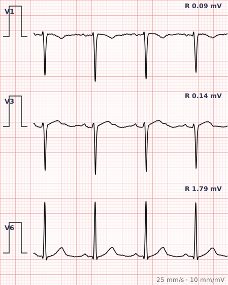
ECG criteria
| Feature | What you see in PRWP |
|---|---|
| Normal pattern | R wave grows steadily from V1 to V5, with the R/S transition usually in V3 or V4. |
| Poor progression | R wave in V3 under 3 mm, or a transition delayed to V5–V6. |
| Reversed progression | R wave amplitude actually decreases from V1 to V4 — more suggestive of pathology. |
| Q waves | Their presence points towards previous anterior infarction rather than a benign cause. |
| ST-T | Concurrent anterior T inversion strengthens the case for previous infarction. |
| Axis | Marked left axis deviation from fascicular block can produce it without any anterior scar. |
How to spot it
- Scan V1 through V6 and watch the R wave height. It should grow steadily.
- Measure the R wave in V3. Under 3 mm meets the usual definition.
- Note where the R/S transition occurs. Normally V3 or V4; later than V5 is delayed.
- Look for pathological Q waves in the anterior leads, which point towards previous infarction.
- Check the chest lead placement. Leads placed too high are the most common cause of the appearance.
- Consider the alternatives before reporting: LVH, left anterior fascicular block, LBBB, chronic lung disease and body habitus all produce it.
What it gets confused with
| Looks like | How to tell them apart |
|---|---|
| Anterior myocardial infarction | Pathological Q waves in the anterior leads, often with T inversion. The diagnosis that matters. |
| Lead misplacement | Chest leads placed too high. Repeating the ECG with careful placement resolves it — the first thing to check. |
| Left ventricular hypertrophy | Voltage criteria met; the tall left-sided voltages shift the transition. |
| Left anterior fascicular block | Marked left axis deviation with rS inferiorly. |
| Left bundle branch block | Wide QRS with a broad R in V6; anterior R waves are lost as part of the block. |
| Chronic lung disease | Low voltages throughout, right axis deviation, and often right atrial enlargement. |
| Body habitus | Obesity, emphysema or a large chest reduce anterior voltages generally. |
Traps
- Lead placement is the most common cause and the first thing to check. Chest electrodes placed an intercostal space too high routinely produce poor R wave progression that vanishes when the ECG is repeated correctly.
- Poor R wave progression is non-specific. On its own it should not be reported as evidence of previous anterior infarction.
- Look for Q waves and T wave changes. Poor progression with anterior Q waves is a much stronger signal than poor progression alone.
- Compare with any previous ECG. A change over time carries far more weight than a single tracing.
- The finding is common in healthy people, particularly women and those with a larger chest, and needs interpreting with the clinical picture rather than in isolation.
Why it happens
Normally the R wave grows across the chest leads as the electrodes move closer to the left ventricle and the depolarisation vector turns towards them. That progression is lost when anterior myocardium is scarred and no longer generates a vector, when the activation sequence is abnormal, when the heart sits differently within the chest, or simply when the electrodes are not where they are assumed to be.
Why it matters
Poor R wave progression is a common and frequently over-interpreted finding. Its importance lies almost entirely in context: with anterior Q waves and T wave inversion it supports previous anterior infarction, while in isolation it is more often explained by lead placement, body habitus or an unrelated conduction abnormality.
Questions
What is poor R wave progression?
Failure of the R wave to grow normally across the precordial leads, usually defined as an R wave in lead V3 measuring less than 3 millimetres, or an R/S transition delayed beyond lead V4.
Does poor R wave progression mean a previous heart attack?
Not on its own. It has many benign causes including lead misplacement, body habitus and chronic lung disease. It supports previous anterior infarction when accompanied by pathological Q waves and anterior T wave inversion.
What should I check first when I see poor R wave progression?
Chest lead placement. Electrodes positioned an intercostal space too high are the most common cause, and repeating the ECG with correct placement resolves the appearance.
Reading about poor r wave progression is not the same as calling it on a tracing you have never seen.
Practise on real cases in ECG Pro