2nd Degree AV Block on the ECG
Second-degree atrioventricular block is intermittent failure of conduction between atria and ventricles, in which some P waves are followed by a QRS complex and others are not, subdivided into Mobitz I with progressive PR lengthening and Mobitz II with a fixed PR.
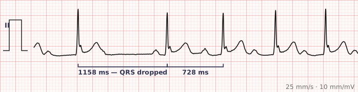
ECG criteria
| Feature | What you see in 2nd Degree AV Block |
|---|---|
| Conduction | Intermittent. Some P waves conduct, some are dropped. More P waves than QRS complexes. |
| Mobitz I (Wenckebach) | PR lengthens progressively over successive beats until one P wave fails to conduct, then the cycle restarts. R-R intervals shorten before the drop. |
| Mobitz II | PR is constant, then a P wave suddenly fails to conduct with no warning. Often accompanied by a wide QRS. |
| 2:1 block | Every other P wave conducts. Cannot be classified as I or II from a single strip, because there are no consecutive conducted beats to compare. |
| QRS | Usually narrow in Mobitz I (block within the node). Often wide in Mobitz II (block below the node). |
| Rate | Ventricular rate is a fraction of the atrial rate, depending on the conduction ratio. |
How to spot it
- Confirm there are more P waves than QRS complexes. That establishes second-degree block.
- March out the P waves. They should be regular — the atria keep going normally.
- Measure the PR interval on each conducted beat in sequence. Progressive lengthening before the dropped beat means Mobitz I.
- If every conducted PR is identical and a beat then drops without warning, that is Mobitz II.
- Look at the R-R intervals in Mobitz I: they characteristically shorten before the pause, which is counter-intuitive and a useful confirmatory sign.
- Check the QRS width. Narrow favours nodal block (Mobitz I); wide favours infranodal block (Mobitz II) and is more concerning.
What it gets confused with
| Looks like | How to tell them apart |
|---|---|
| Blocked PAC | An early, abnormally shaped P wave fails to conduct. In AV block the non-conducted P arrives on time. |
| Sinus pause or arrest | No P wave at all during the pause. In AV block a P wave is present but not conducted. |
| Third-degree AV block | No P wave conducts at all, and PR intervals vary randomly. In second-degree block some P waves clearly do conduct. |
| First-degree AV block | PR is long but nothing is ever dropped. |
| Atrial flutter with block | Flutter waves at about 300 rather than discrete P waves at a normal atrial rate. |
Traps
- Non-conducted P waves hide in T waves. A T wave that looks taller, notched or peaked compared with the others usually has a P buried in it. Comparing T wave shapes across the strip is the single most useful habit here.
- The distinction matters clinically. Mobitz I is usually benign and often vagal; Mobitz II reflects disease below the AV node, progresses to complete block, and generally means a pacemaker.
- 2:1 block cannot be labelled Mobitz I or II from one strip — there are never two consecutive conducted beats to compare PR intervals. Say "2:1 AV block" and look at a longer recording.
- In Wenckebach the R-R interval shortens before the dropped beat, which surprises people who expect it to lengthen. The PR increment gets smaller each beat even as the absolute PR grows.
- A group of beats followed by a pause — "grouped beating" — should always trigger a careful measurement of every PR interval in the group.
Why it happens
In Mobitz I the AV node itself fatigues: each successive impulse arrives while the node is progressively less recovered, so conduction takes longer each beat until one impulse fails entirely, after which the node recovers fully and the cycle restarts. In Mobitz II the block is below the node, in the His bundle or bundle branches, where conduction is all-or-nothing — so it either works normally or fails abruptly, with no warning.
Why it matters
The two types behave very differently. Mobitz I is frequently vagal, often seen in athletes or during sleep, and rarely progresses. Mobitz II reflects structural disease of the infranodal conduction system, is unpredictable, and can deteriorate to complete heart block without warning — which is why it usually leads to pacing regardless of symptoms.
Questions
What is the difference between Mobitz I and Mobitz II?
In Mobitz I the PR interval lengthens progressively over consecutive beats until one P wave fails to conduct. In Mobitz II the PR interval stays constant and a P wave drops out suddenly. Mobitz I is usually within the AV node and benign; Mobitz II is below it and usually requires pacing.
Why can 2:1 AV block not be classified?
Classifying requires comparing PR intervals on consecutive conducted beats. In 2:1 block every other beat is dropped, so no two conducted beats are adjacent and progressive PR lengthening cannot be assessed. A longer rhythm strip that captures a different conduction ratio is needed.
Why do R-R intervals shorten before the dropped beat in Wenckebach?
Although the PR interval lengthens each beat, the size of each increment gets smaller. Because the R-R interval reflects the change in PR rather than its absolute value, decreasing increments mean progressively shorter R-R intervals.
How do I find a non-conducted P wave?
Compare T wave shapes across the strip. A non-conducted P wave superimposed on a T wave makes it taller, notched or differently shaped than its neighbours. Marching out the P waves at a fixed interval also shows where the hidden one must be.
Reading about 2nd degree av block is not the same as calling it on a tracing you have never seen.
Practise on real cases in ECG Pro