Atrial Flutter on the ECG
Atrial flutter is a supraventricular arrhythmia driven by a re-entry circuit within the atria, producing regular sawtooth flutter waves at approximately 300 per minute and a ventricular rate that is usually a simple fraction of that.
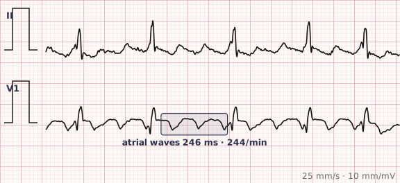
ECG criteria
| Feature | What you see in Atrial Flutter |
|---|---|
| Rate | Atrial rate 250–350, typically 300. Ventricular rate is a fraction of it: 150 with 2:1 conduction, 100 with 3:1, 75 with 4:1. |
| Rhythm | Regular when the conduction ratio is fixed; irregular when the ratio varies. |
| P wave | Replaced by flutter waves — a continuous sawtooth with no isoelectric baseline between them. Best seen in II, III, aVF and V1. |
| PR interval | Not applicable. Describe the conduction ratio instead. |
| QRS | Narrow, under 120 ms, unless there is bundle branch block or aberrancy. |
| ST-T | Hard to assess — flutter waves superimpose on the ST segment and mimic depression. |
How to spot it
- Any regular narrow-complex tachycardia at about 150 should make you look for flutter before anything else.
- Go to leads II, III and aVF and look at the baseline between QRS complexes. Flutter gives a continuous sawtooth with no flat segment; in V1 the waves are often discrete and upright.
- Count the atrial rate. If it is close to 300, and the ventricular rate is 150, 100 or 75, the diagnosis is nearly made.
- Work out the conduction ratio by counting flutter waves per QRS. Report it — "flutter with 2:1 block" says more than "flutter".
- If flutter waves are hidden inside QRS complexes and T waves, a vagal manoeuvre or adenosine transiently slows conduction and exposes them.
What it gets confused with
| Looks like | How to tell them apart |
|---|---|
| Sinus tachycardia | Discrete P waves with a flat isoelectric baseline between them, and a rate that drifts rather than sitting at exactly 150. |
| Supraventricular tachycardia | No flutter waves; rate often above 150, and it terminates abruptly with adenosine rather than transiently slowing. |
| Atrial fibrillation | Chaotic and irregular with no organised atrial waves. Flutter is organised, even when its conduction ratio varies. |
| Ventricular tachycardia | Wide QRS. Flutter with aberrancy can be wide too — look for flutter waves marching through the complexes. |
Traps
- 2:1 flutter is the most commonly missed arrhythmia on the ECG. Every other flutter wave hides inside the QRS or T wave, leaving what looks like a plain regular tachycardia at 150.
- Flutter waves distort the ST segment and T wave, so ischaemia cannot be reliably assessed until the rhythm is controlled.
- A ventricular rate that is almost exactly 150, 100 or 75 is a strong hint. Rates land on those numbers because 300 divides by small whole numbers.
- Adenosine does not usually cardiovert flutter. It slows AV conduction long enough to reveal the flutter waves, which is diagnostic rather than therapeutic — warn the patient first.
Why it happens
A macro-re-entrant circuit, most often running counter-clockwise around the tricuspid annulus in the right atrium and passing through the cavotricuspid isthmus, depolarises the atria in a continuous loop at about 300 per minute. The AV node cannot conduct every impulse, so it blocks a fixed proportion, which is why the ventricular rate is a clean fraction of the atrial rate.
Why it matters
Atrial flutter carries a stroke risk comparable to atrial fibrillation and frequently coexists with it, so anticoagulation is assessed the same way. Because the circuit is anatomically defined, catheter ablation of the cavotricuspid isthmus is highly effective — which makes recognising flutter rather than assuming AF worth the effort.
Questions
Why is the ventricular rate in atrial flutter so often exactly 150?
The atrial rate is close to 300 and the AV node commonly conducts every second impulse. Half of 300 is 150. The same arithmetic produces 100 with 3:1 conduction and 75 with 4:1.
Where do I look for flutter waves?
Leads II, III and aVF show the classic sawtooth, because the usual circuit runs in the inferior part of the right atrium. Lead V1 often shows discrete upright flutter waves that are easier to count.
How do I reveal flutter waves that are hidden?
Slow AV conduction transiently with a vagal manoeuvre or adenosine. The ventricular rate drops for a few seconds and the underlying flutter waves become visible. This is a diagnostic manoeuvre, not a treatment.
Does atrial flutter need anticoagulation like atrial fibrillation?
Stroke risk in flutter is treated as comparable to atrial fibrillation, and the two frequently coexist in the same patient, so risk assessment follows the same approach.
Reading about atrial flutter is not the same as calling it on a tracing you have never seen.
Practise on real cases in ECG Pro