1st Degree AV Block on the ECG
First-degree atrioventricular block is a delay in conduction between the atria and ventricles, defined by a PR interval longer than 200 milliseconds in which every P wave is still followed by a QRS complex.
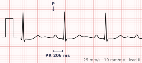
ECG criteria
| Feature | What you see in 1st Degree AV Block |
|---|---|
| PR interval | Over 200 ms (one large square), and constant from beat to beat. |
| Conduction | 1:1 — every P wave conducts. Nothing is dropped. This is the entire distinction from second-degree block. |
| Rate | Determined by the underlying rhythm, usually sinus. |
| Rhythm | Regular. |
| P wave | Normal sinus P before every QRS. |
| QRS | Usually narrow. A wide QRS alongside a long PR suggests disease below the AV node as well. |
How to spot it
- Measure the PR interval from the start of the P wave to the start of the QRS. Over one large square (200 ms) is prolonged.
- Confirm the PR is the same on every beat. A PR that lengthens progressively is Wenckebach, not first-degree block.
- Confirm every P wave is followed by a QRS. A single dropped beat makes it second-degree block.
- Check the QRS width. A narrow QRS points to delay in the AV node itself; a wide QRS raises the possibility of additional disease in the bundle branches.
- Look at a very long PR carefully — above about 300 ms the P wave can fall on the preceding T wave and be missed entirely.
What it gets confused with
| Looks like | How to tell them apart |
|---|---|
| Mobitz I (Wenckebach) | PR lengthens progressively until a beat is dropped. In first-degree block the PR is long but fixed. |
| Complete heart block with coincidental timing | P waves and QRS complexes are dissociated, so the PR varies. First-degree block has a fixed relationship. |
| Junctional rhythm with retrograde P | P waves after or buried in the QRS rather than genuinely preceding it. |
| Blocked PAC | A pause with an early hidden P wave, rather than uniform PR prolongation. |
Traps
- "Block" is a misnomer here — nothing is blocked. Every impulse gets through, just slowly. It is a conduction delay.
- A markedly long PR can push the P wave into the preceding T wave, making it look as though there is no P at all. Compare T wave shapes across the strip.
- Isolated first-degree block in an asymptomatic person is usually benign and needs nothing.
- First-degree block combined with bifascicular block is a different matter, and has traditionally raised concern about progression to complete block.
- Drugs are a common cause — beta blockers, calcium channel blockers, digoxin and amiodarone all prolong AV nodal conduction.
Why it happens
Conduction through the AV node is slowed, usually by increased vagal tone, drugs that act on the node, or age-related fibrosis. The impulse still reaches the ventricles every time; it simply takes longer to get through, which lengthens the interval between atrial and ventricular depolarisation.
Why it matters
On its own, first-degree AV block is generally benign and requires no treatment. It becomes more interesting when it is new, when the PR is very long, when it accompanies bundle branch block, or when a rate-slowing drug is being started — situations where it may be the first sign of more extensive conduction disease.
Questions
What PR interval defines first-degree AV block?
Longer than 200 milliseconds — one large square on standard paper — measured from the beginning of the P wave to the beginning of the QRS complex, with every P wave still conducting.
Is first-degree AV block dangerous?
In isolation it is usually benign and needs no treatment. It matters more when it is new, when it accompanies bundle branch block, or when it appears while a rate-slowing drug is being introduced.
Why is it called a block if nothing is blocked?
The name is historical and inaccurate. Every atrial impulse reaches the ventricles; conduction is merely delayed. It is more accurately described as first-degree AV conduction delay.
Reading about 1st degree av block is not the same as calling it on a tracing you have never seen.
Practise on real cases in ECG Pro