Sinus Rhythm on the ECG
Sinus rhythm is the normal cardiac rhythm, in which each beat begins in the sinoatrial node and produces an upright P wave in lead II followed by a QRS complex at a rate of 60 to 100 beats per minute.
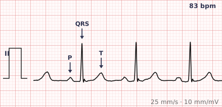
ECG criteria
| Feature | What you see in Sinus Rhythm |
|---|---|
| Rate | 60–100 bpm. Below 60 is sinus bradycardia, above 100 is sinus tachycardia — both are still sinus rhythm. |
| Rhythm | Regular. Beat-to-beat variation under about 10% of the R-R interval. |
| P wave | Present before every QRS. Upright in II, III and aVF; inverted in aVR. Same shape every beat. |
| PR interval | 120–200 ms, and constant. |
| QRS | Under 120 ms, unless there is co-existing bundle branch block. |
| ST-T | Isoelectric ST segment; T wave upright in most leads and concordant with the QRS. |
How to spot it
- Look at lead II. If the P wave is upright there and inverted in aVR, the impulse is travelling from the sinus node down towards the apex — that is the definition of sinus.
- Check every QRS has a P in front of it, and every P is followed by a QRS. One-to-one, both ways.
- March out the R-R intervals. Sinus rhythm is regular, though a young patient may show clear respiratory variation.
- Measure the PR. It must be 120–200 ms and identical on every beat.
- Confirm the rate is 60–100. Outside that range the rhythm is still sinus, just fast or slow — say which.
What it gets confused with
| Looks like | How to tell them apart |
|---|---|
| Ectopic atrial rhythm | Regular with 1:1 P waves, but the P is inverted in II or abnormally shaped — the focus is not the sinus node. |
| Atrial fibrillation | No P waves at all and an irregularly irregular ventricular response. |
| Sinus arrhythmia | Sinus P waves with rate varying by breath — normal in the young, not a disease. |
| Wandering atrial pacemaker | At least three different P wave shapes as the pacemaker site drifts. |
| Junctional rhythm | Regular and narrow, but P waves are absent, inverted or buried, and the rate is typically 40–60. |
Traps
- Sinus rhythm is not the same as a normal ECG. A tracing can be in sinus rhythm and still show infarction, hypertrophy or bundle branch block. The rhythm is one line of the report, not the report.
- A T wave with a P wave riding on it can look like a single deflection at fast rates. Slow down and find the P before calling anything junctional.
- Lead II is the lead for P wave axis. Judging P morphology from V1 alone leads to trouble — the P is often biphasic there normally.
- Respiratory variation in a young person is sinus arrhythmia, which is normal. Do not report it as an irregular rhythm requiring investigation.
Why it happens
The sinoatrial node, in the high right atrium, depolarises spontaneously faster than any other cardiac tissue and so sets the pace. Depolarisation spreads down and leftward across the atria towards the AV node, which is why the resulting P wave points towards lead II and away from aVR.
Why it matters
Sinus rhythm is the reference point for the whole tracing. Establishing it first tells you the atria and the AV node are conducting normally, which narrows everything you look at afterwards. It is also the rhythm most treatments for other arrhythmias are trying to restore.
Questions
Is sinus rhythm always normal?
The rhythm is normal, but the ECG may not be. Sinus rhythm only describes where the beat starts and how it reaches the ventricles. Ischaemia, hypertrophy, bundle branch block and infarction all occur in sinus rhythm.
What rate counts as sinus rhythm?
Conventionally 60 to 100 beats per minute. Outside that range the rhythm is still sinus — it is called sinus bradycardia below 60 and sinus tachycardia above 100.
How do I prove the P wave is from the sinus node?
Check its direction. A sinus P wave is upright in leads II, III and aVF and inverted in aVR, because the impulse travels down and to the left. A P wave inverted in II did not start in the sinus node.
References
Reading about sinus rhythm is not the same as calling it on a tracing you have never seen.
Practise on real cases in ECG Pro