Fusion Beats on the ECG
A fusion beat is a QRS complex produced when a supraventricular impulse and a ventricular impulse depolarise the ventricles simultaneously, giving a complex whose shape is intermediate between a normal conducted beat and a purely ventricular one.
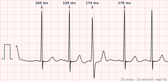
ECG criteria
| Feature | What you see in Fusion Beats |
|---|---|
| Timing | Occurs when an ectopic ventricular impulse coincides with a conducted supraventricular beat. |
| QRS | Intermediate in shape and width — narrower than the ventricular beat, wider than the sinus beat, and clearly a hybrid of the two. |
| P wave | A P wave usually precedes the fusion beat, at a PR interval slightly shorter than normal. |
| Context | Seen with PVCs, accelerated idioventricular rhythm, ventricular tachycardia and paced rhythms. |
| Significance | During a broad-complex tachycardia, a fusion beat is strong evidence for ventricular tachycardia. |
| ST-T | Also intermediate, following the hybrid QRS. |
How to spot it
- Identify the two beat shapes on the tracing: the normal conducted QRS and the ventricular one.
- Look for a complex that is neither — a shape that sits visually between the two.
- Check for a preceding P wave with a slightly shortened PR, which shows the supraventricular impulse arrived and contributed.
- In a broad-complex tachycardia, hunt deliberately for fusion and capture beats. They are uncommon but close to diagnostic.
- In a paced rhythm, a fusion beat means the patient's own conduction and the pacemaker fired together — usually normal behaviour, not malfunction.
What it gets confused with
| Looks like | How to tell them apart |
|---|---|
| Premature ventricular contraction | A PVC is fully ventricular, so it is wide and bizarre throughout with no intermediate shape. |
| Capture beat | A fully conducted normal-looking beat during a ventricular rhythm, rather than a hybrid. Also strong evidence for VT. |
| Aberrantly conducted beat | Follows a typical bundle branch block pattern rather than an intermediate shape. |
| Pacemaker pseudofusion | A pacing spike lands on an intrinsic beat without contributing to it, so the QRS shape is unchanged. |
Traps
- Fusion beats during a broad-complex tachycardia are one of the strongest pieces of evidence for ventricular tachycardia. They are easy to miss because they are single beats — scan the whole strip for a complex that looks intermediate.
- Fusion in a paced rhythm is normal, not a fault. It simply means intrinsic conduction and pacing coincided.
- A fusion beat can be mistaken for an artefact or a poorly conducted beat. Its shape being consistently intermediate between two known morphologies is the giveaway.
- Fusion beats occur in accelerated idioventricular rhythm, where the ventricular and sinus rates are close and the two compete for the ventricle.
Why it happens
When a supraventricular impulse descends the conduction system at the same moment a ventricular focus fires, each depolarises part of the ventricular myocardium. The resulting QRS is the electrical sum of the two activation wavefronts, so its shape lies between the two pure forms in proportion to how much of the ventricle each contributed.
Why it matters
Fusion beats are not a problem in themselves; their value is entirely diagnostic. Finding one during a broad-complex tachycardia demonstrates that a supraventricular impulse reached the ventricle independently, which means the tachycardia is arising below the atria — powerful evidence for ventricular tachycardia at a moment when that distinction matters most.
Questions
What causes a fusion beat?
Two activation wavefronts arriving at the ventricles at the same time — one conducted normally from above, one from a ventricular focus. The QRS is the electrical sum of both, so its shape is intermediate.
Why do fusion beats suggest ventricular tachycardia?
A fusion beat proves a supraventricular impulse reached the ventricle independently of the tachycardia, which means the tachycardia is not itself supraventricular. Along with capture beats and AV dissociation, it is among the most reliable signs of VT.
Are fusion beats in a paced rhythm a problem?
No. They mean the patient's own conduction and the pacemaker happened to activate the ventricle together, which is expected behaviour when intrinsic rate and paced rate are similar.
Reading about fusion beats is not the same as calling it on a tracing you have never seen.
Practise on real cases in ECG Pro